Sijie Zhou

and 5 more

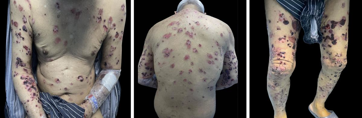
Manuscript category: Letters to the EditorTitle: New-onset bullous pemphigoid after SARS-CoV-2 infectionKeywords: COVID-19, Bullous pemphigoid, SARS-CoV-2Word count: 451 wordsFigure count: 2Table count: 0Sijie Zhou1, Xue Wang1, Lizhi Ma1, Jiaming Fan1, Xinyun Tang1, Peimei Zhou1*1 Department of Dermatovenereology, Chengdu Second People’s Hospital, Qingyun Street, Chengdu, 610041, China.Correspondence to: Peimei Zhou, M.D., Ph.D.Department of Dermatovenereology, Chengdu Second People’s Hospital, Qingyun Street, Chengdu, 610041, China.Tel: +86 18908176315; E-mail: [email protected] sources: NoneConflict of interest disclosures: All authors declare no conflicts of interest.Data availability statement: Data sharing does not apply to this article as no new data were created or analysed in this study.Ethics statement: The patient consented to publish this information. A 78-year-old male patient with a history of diabetes, hypertension, and renal insufficiency was diagnosed with bullous pemphigoid (BP) after contracting coronavirus disease 2019 (COVID-19). The patient had a positive nasopharyngeal swab for severe acute respiratory syndrome coronavirus 2 (SARS-CoV-2) in late December 2022 and was hospitalised for two to three weeks. After discharge, the patient developed erythema and blisters on both hands with itching that gradually involved the whole body. The patient presented to our dermatology department on 3 March 2023.At presentation to the dermatology department, the patient had tense blisters covering the entire body, some of which were breaking down, leaving a small patchy vesicular surface with a yellowish exudate (Fig. 1). Routine blood, procalcitonin, and C-reactive protein tests suggested infection, and a chest computed tomography exam indicated viral pneumonia, consistent with SARS-CoV-2 infection. In addition, the patient’s BP180 antibody levels were >150 U/mL (range: positive >20 U/mL), and rickle cell desmosome antibodies were negative. Furthermore, direct immunofluorescence showed linear basal deposition of immunoglobin G and C3 (uncertain). Finally, histological analyses of an incisional cutaneous biopsy obtained from the patient’s left lower extremity showed the formation of subepidermal blisters containing a few eosinophils, lymphocytes and fibrin, and some epidermal basal cells next to the blisters were edematous, liquefied and deformed.(Fig. 2). These findings were consistent with a BP diagnosis. The patient’s condition did not resolve after conventional treatment with glycyrrhizin, a Chinese herbal extract compound, and other anti-inflammatory treatments; therefore, dupilumab (600 mg initially, then 300 mg every two weeks after) was subcutaneously administered. The symptoms were relieved after two weeks.An increasing number of studies are reporting cutaneous manifestations after COVID-19. For instance, nonspecific skin symptoms, including urticarial lesions, chilblain-like lesions, vesicular eruptions, maculopapular rashes, and livedo, have been reported1,2. We identified no other reports of COVID-19-induced BP but did find a description of BP after SARS-CoV-2 vaccination3. The vaccine mimics antigens to induce specific immune responses in the body, similar to those produced by humans infected with novel coronaviruses, which can cause autoimmune symptoms4. Therefore, our case was likely associated with a novel coronavirus infection.BP is an autoimmune, subepidermal blistering disease occurring in elderly individuals. The exact pathogenesis of BP is unknown, but several factors, such as drugs, thermal or electrical burns, surgical procedures, trauma, ultraviolet irradiation, radiotherapy, chemical preparations, transplants, and infections, may induce or exacerbate BP5. Yet, viral infection-induced BP has rarely been considered. This may be because viruses, as pathogens, can induce cross-reactive autoantibodies sharing epitopes with host cells. Additionally, the virus can directly infect keratinocytes, induce the expression of hidden epitopes, modify existing epitopes, or insert envelope fragments into cells to produce new antigens, which could be responsible for BP development6,7.

Yihang Xie

and 4 more

New-onset pemphigus after COVID-19Yihang Xie1 Mei Yang1 Peimei Zhou1* Jiaming Fan1 Sijie Zhou11.Department of Dermatovenereology, Chengdu Second People’s Hospital, Chengdu, China* Corresponding Author: [email protected] head: pemphigus after COVID‐19The category of the article: LetterKeywords: COVID‐19, pemphigusManuscript word count: 690 wordsThe number of figures: 2The number of tables: 0The number of references: 8Correspondence to: Peimei Zhou, M.D, Ph.D., Department of Dermatovenereology, Chengdu Second People’s Hospital, Qingyun Street, Chengdu, 610041, China.Tel: +86 18908176315; E-mail:[email protected] conflict of interest statement: Y. Xie, and my co-authors have no conflict of interest to declare.Ethics statement: The patient has consented to publish this information.Data availability statement: Data sharing does not apply to this article as no new data were created or analyzed in this study.Funding sources: noneDear Editor,Cutaneous manifestations of coronavirus disease (COVID-19), the disease caused by severe acute respiratory syndrome coronavirus 2 (SARS-CoV-2; family Coronaviridae, genus Betacoronavirus, subgenusSarbecovirus ), have been increasingly reported. SARS-CoV-2 infection is multisystemic and leads to potentially detrimental effects on various organs. Maculopapular, urticarial, vesicular, livedoid, and Chilblain-like lesions (CBLL) have been commonly reported to be associated with COVID-191. Here, we encountered an intriguing case of pemphigus that developed after COVID-19 infection.A 73-year-old male presented with a 42-day history of pruritic flaccid blisters that arose on the trunk and both upper limbs on normal and erythematous skin. Cutaneous lesions started 3 days after the positive reverse transcription polymerase chain reaction (RT-PCR) test diagnostic for SARS-CoV-2. He denied any history of systemic diseases, medication, and medicine or food allergies, and had not used any medication before symptom onset. The patient had first been diagnosed with allergic dermatitis caused by COVID-19 at another hospital and was prescribed oral prednisone (8 mg once daily for 4 days). The patient reported no new blisters, but the erythema did not fade; therefore, he visited our hospital. Physical examination revealed cutaneous lesions on the trunk and both upper limbs without mucosal involvement and scattered superficial blisters that developed into crusted erosions on an erythematous base(Figure1 A-D). Laboratory examination revealed normal white cell count (8.63 Ö109/L; normal 3.5-9.5 Ö109/L) with eosinophilia (6%; normal 0.5%-5.0%). Desmoglein (Dsg) 1 antibody levels were > 150 U/mL (positive: > 20), while Dsg3, BP(bullous pemphigoid)180, and BP230 antibody levels were within normal ranges. Other laboratory tests including RT-PCR targeting SARS-CoV-2, immunoglobulin, erythrocyte sedimentation rate, the spectrum of antinuclear antibodies, and T-spot were negative or normal. Chest and abdominal computed tomography revealed chronic inflammatory changes but no obvious tumors. Histological analysis of an incisional cutaneous biopsy taken from the patient’s abdomen showed subcorneal blister formation, acantholytic cells within the blister, and marked spongiotic edema in the spongiosa layer that had mixed inflammatory infiltrate with eosinophils, leukomonocytes, and neutrophils(Figure2A). Direct immunofluorescence (DIF) showed deposition of intracellular IgG and C3 in subepidermal 2/3 interspinous cells, though was negative for IgA and IgM, confirming pemphigus(Figure2B,C). Considering the good response to hormone treatment, the patient continued oral prednisone at 8 mg once daily along with the use of topical corticosteroids. Symptoms were completely absent after 3 weeks(Figure1 E-F).An increasing number of studies on cutaneous manifestations of COVID-19 have been reported; however, knowledge is still lacking on the common skin manifestations of this disease. Nonspecific cutaneous manifestations due to SARS-CoV-2 infection have also been reported, such as immune thrombocytopenic purpura (ITP), dengue-like exanthem, pityriasis rosea-like eruptions, acral ischemia, mucositis, dusky lesions, and bullae2.3.4. We searched all relevant articles and found only two cases of pemphigus vulgaris induced by COVID-19. In the case presented here, we realized that COVID-19 may be responsible for the rash eruption, possibly due to an inflammatory reaction5. The onset time of the rash was similar to that in the cases of pemphigus previously reported by De Medeiros5 and Mohaghegh F6 (within 1.5 months). In our case, although direct immunofluorescence showed subepidermal 2/3 deposition, we still diagnosed pemphigus foliaceus in combination with the pathological presentation, indirect immunofluorescence, and good treatment outcome. We speculate that the reason why direct immunofluorescence showed subepidermal 2/3 deposition may be the marked sponge edema of the epidermis, which may lead to a discontinuity of acantholysis, resulting in leakage of Dsg1 into the deeper epidermis.Pemphigus is defined as a group of rare mucocutaneous autoimmune diseases. Its etiology is unknown, though there are studies on autoimmune etiology which is believed to be related to stimulation by certain drugs, ultraviolet radiation, and malignant tumors; these induce autoimmune reactions by making the adhesive substances between the spiny cell layers become autoantigens7. It is rarely considered, however, that viral infections might cause pemphigus. The ability of SARS-CoV-2 to induce a hyper-stimulated immune state was discovered at the beginning of the pandemic8. As an instrumental trigger of autoimmunity, SARS-CoV-2 infection could be a trigger for autoimmune reactions, possibly through more than one mechanism. Because of this, all factors should be considered in any patient presenting with new-onset or exacerbating cutaneous reactions.