Hoda Abdelgawad

and 3 more

14-years old asymptomatic boy with holosystolic murmur heard over the lower left sternal border. Zoomed apical 5 chamber view revealed a perimembranous ventricular septal defect (VSD) which was closed by an aneurysmal leaflet of the tricuspid valve (TV) (Figure A) .Color Doppler across the defect showed left to right shunt (Figure B) Short axis view at the level of the aortic valve showed that VSD was extending from 10 to 11 o’clock . (Figure C). Injection of bubbles showed negative contrast inside the right ventricle indicating incomplete defect closure. (Figure D) 3D Transesophageal Echocardiography (TOE) with zoomed mode showed the entry point from the left ventricular side and exit point from the right ventricular side.(Figure E) En-face viewing of the TV from the right ventricular perspective showed an aneurysm-like pouch formed by the anterior leaflet (Figure F) The left atrial and ventricular size was normal and the shunt ratio was 1.3 so he was treated conservatively. Ventricular septal defect is the most common congenital anomaly.The incidence of spontaneous closure is still undetermined .Different mechanisms for anatomical closure have been proposed such as the adherence of the septal leaflet and/or the anterior leaflet of the tricuspid valve to the margins of the defect forming an aneurysm-like pouch that maybe mistaken for an aneurysm of the membranous septum. Echocardiography is the gold standard for evaluation of the site , size and type of VSD and its relation to the tricuspid valve. Recently, transcatheter closure is feasible and efficacious in properly selected patients.

Fatma Elkafrawy

and 3 more

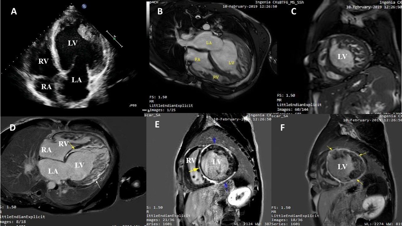
Myocardial non-compaction (NC) is a rare genetically heterogeneous cardiomyopathy which is characterized by excessively prominent ventricular trabeculations with deep intertrabecular recesses. It is commonly believed to be due to an intrauterine arrest of the endomyocardial morphogenesis. Left ventricle (LV) is the usual site of affection, but very rarely right ventricular (RV) or both ventricles may be involved. It can be isolated or associated with other cardiac diseases or neuromuscular disorders. It has a wide spectrum of clinical manifestations that range from asymptomatic status, progressive ventricular dysfunction, arrhythmias and congestive heart failure that have been found to be associated with myocardial fibrosis. Cardiac Magnetic resonance (CMR) is the gold standard non-invasive modality for the diagnosis of cardiomyopathies. In NC cases, late gadolinium enhancement (LGE) of the trabeculated layer or mild fibrosis of the compacted layer was previously described in the literature, however extensive fibrosis in the compacted layer only with no fibrosis at the trabeculated layer was not described before. We report a male patient presented with heart failure symptoms, echocardiogram showed severe LV and RV systolic dysfunction with prominent bi-ventricular trabeculations. CMR confirmed the diagnosis of bi-ventricular non-compaction with peculiar pattern extensive fibrosis of the compacted layer of LV in LGE. To the best of our knowledge, our case is the first case to report extensive fibrosis in the compacted layer only with no fibrosis at the trabeculated layer.