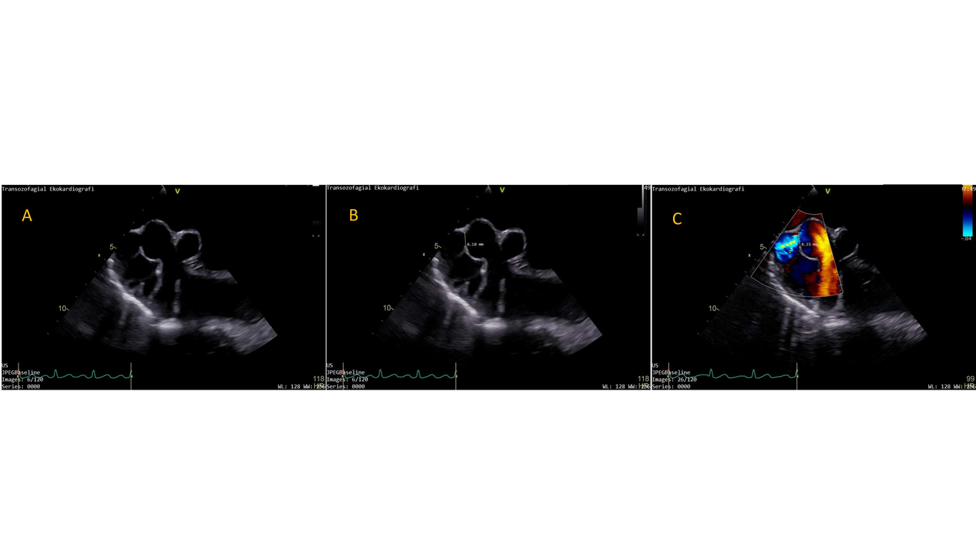
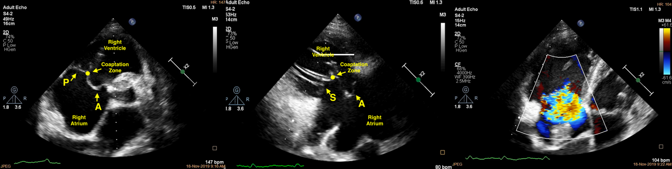
Background: Coronary artery fistula (CAF) is a congenital connection between a coronary artery and cardiac chambers, or a vessel bypassing a capillary system. The clinical presentation of congenital CAF varies, depending on its size and the draining chamber. Case presentation: A 23-year-old patient without a prior medical history consulted to cardiology clinic with a referral diagnosis of interatrial septal aneurysm and defect. Transthoracic echocardiography revealed a dilated right coronary artery to a sac into the right atrium with dilated right side of the heart. Transesophageal echocardiography provided the calculation of the narrowest part of the fistula where the closure device was planned to be deployed, and two sequential sacs of the fistula on its way to the right atrium. Cardiac computed tomography demonstrated the aneurysmatic dilatation of the proximal portion of the right coronary artery (CA) was 14*20 mm and the narrowest exit of the CAF as 8*8.3 mm width. transcatheter closure of an isolated enormous CAF was decided and surgery left to be reserved for CAF with failed percutaneous closure. Conclusion: Multi-modality imaging take part in the diagnosis and follow-up of the CAFs for identifying the anatomy/physiology, guiding the intervention, supplementary hemodynamic information, assessing the anatomical relationship and ischemia.